
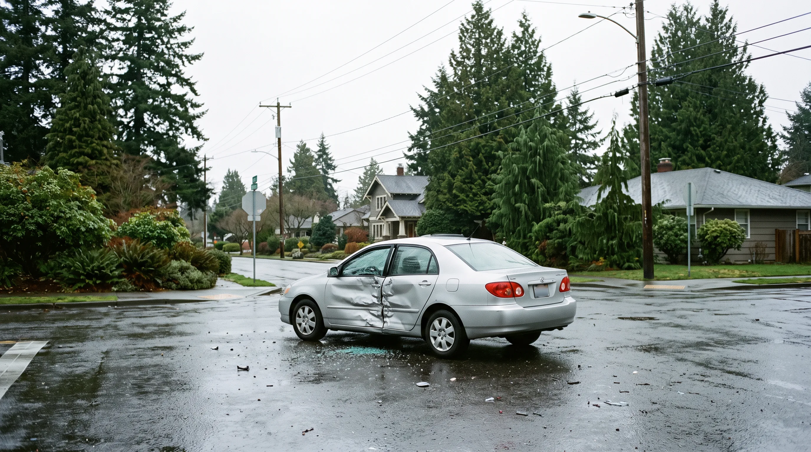
A 41-year-old project manager came into our clinic about three days after her car was struck on the driver’s side at a West Linn intersection. She had already been to the emergency room the night of the crash, where X-rays came back negative for fractures. She was discharged with a prescription for muscle relaxants and instructions to follow up with her primary care physician. By the time she walked through our door, she described her pain as “a six out of ten and getting worse.”
Her situation is one I see regularly. Side impact car accident injuries often present with a deceptively clean initial picture, only to become significantly more complex over the following days and weeks. This case study walks through her mechanism of injury, our diagnostic approach, her treatment course, and the functional milestones that marked her recovery. Names and identifying details have been changed; this is an anonymized composite that protects patient privacy.
Why Side Impact Car Accident Injuries Are Different
Rear-end collisions get most of the clinical and legal attention, but T-bone crashes carry a different injury profile that clinicians need to understand and that patients deserve to know about.
In a rear-end collision, the head and neck are thrown backward and then forward along a predictable axis, and the cervical spine absorbs that whiplash load over a fraction of a second.
In a side impact collision, the loading is lateral. The torso is pushed hard toward the point of impact while the head and neck lag behind due to inertia. This lateral flexion and rotation combination places simultaneous tensile and compressive forces on structures the cervical spine was not designed to protect in that direction: the contralateral facet joints, the brachial plexus, the shoulder capsule, and the lateral musculature of the neck and mid-back.
Our patient presented with left-sided neck pain, pain across the top of her left shoulder, a persistent headache localized at the base of her skull, and intermittent numbness in her left forearm and two fingers. The finger numbness was the clinical finding that changed our initial assessment the most.
Clinical Assessment and What We Found
Cervical range of motion was significantly restricted in lateral flexion and rotation toward the left. Orthopedic testing was positive for C6 nerve root compression. Tenderness was concentrated over the left cervicothoracic junction and levator scapulae insertion.
I referred her for cervical MRI before beginning any manual treatment. Imaging confirmed a left-sided C5-C6 disc protrusion with mild neuroforaminal narrowing, consistent with the trauma mechanism. There was no cord compression, and her neurological presentation did not meet the threshold for surgical consultation. She was a candidate for conservative chiropractic care.
Finger numbness after a T-bone collision is not just a “pinched nerve” to be managed with stretching. It is a signal that demands imaging before a provider begins loading the cervical spine.
The Treatment Plan and Recovery Timeline
Weeks 1 through 3. With the MRI in hand, we began a gentle, neurologically informed course of care. This included instrument-assisted soft tissue mobilization to the cervicothoracic region, low-load cervical distraction, and progressive therapeutic exercise targeting deep cervical flexor activation. We avoided high-velocity manipulation of the cervical spine during this acute inflammatory phase. Her headaches, which had been daily, began to decrease in frequency by the end of week two.
Weeks 4 through 7. As the acute inflammatory response settled, we introduced gentle cervical mobilization and expanded the exercise program to include scapular stabilization work. The numbness in her forearm and fingers was the slowest symptom to respond, which is typical with nerve root involvement. By week six, she described the numbness as intermittent rather than constant, and reduced from its peak intensity by roughly half.
She also returned to driving during week five, which was a meaningful functional milestone. Before that point, she had been relying on family members for transportation to her appointments and work commitments.
Weeks 8 through 12. We transitioned to a more active rehabilitation model, adding cervical traction, progressive resistance training for the shoulder girdle, and a home exercise program she could continue independently. By week ten, she was working her full schedule without modification. The residual symptom was occasional left forearm tingling with prolonged overhead work, addressed with postural education and ergonomic recommendations.
Discharge and outcome. At week thirteen, cervical range of motion had returned to normal in all planes, pain scores were consistently zero to one, and the neurological symptoms had resolved. She had a home program to manage any recurrence and a sixty-day re-check scheduled.
Documentation and the Insurance Picture
This patient had Oregon PIP (personal injury protection) coverage, which applies regardless of fault and provides at least $15,000 in medical benefits for chiropractic and other qualified care. Her treatment was billed entirely through PIP for the first several months, which kept her out of pocket costs at zero during her recovery.
Throughout her care, we maintained detailed records: the initial examination, MRI referral and report, visit-by-visit progress notes, functional outcome scores, and a narrative summary linking her injury mechanism to her clinical findings. That documentation became part of her personal injury file.
Our role is medical, not legal. We document what we find, what we do, and how the patient responds. We encourage any patient navigating a personal injury claim to work with a qualified personal injury attorney in Oregon for guidance on their case.
If you have been in a T-bone or intersection crash and are dealing with side impact car accident injuries, do not assume a clean ER X-ray means you are fine. The structures most commonly injured in lateral collisions, including cervical discs, facet joints, and nerve roots, do not show up on plain film.
We are easy to reach. That is what we are here for.
Dr. Tristan Santiago Maceiras is a Doctor of Chiropractic at Gentle Care Chiropractic in West Linn, Oregon, specializing in musculoskeletal medicine, MVA recovery, and personal injury care.
Frequently Asked Questions
What makes side impact car accident injuries different from rear-end collision injuries? In a T-bone collision, the force is applied laterally to the occupant, creating a lateral flexion and rotation load on the cervical spine that rear-end crashes do not produce. This places different structures at risk, including the contralateral facet joints, the brachial plexus, and the shoulder capsule, and often leads to injury patterns that develop or worsen over the days following the crash.
How long does T-bone collision recovery typically take? Recovery timelines vary considerably based on the structures involved. Soft tissue injuries without disc or nerve involvement often resolve with four to eight weeks of conservative care. When a cervical disc protrusion with nerve root compression is present, as in this case study, active treatment may run twelve to sixteen weeks, with some residual symptoms persisting longer. Consistent care and active patient participation are the strongest predictors of outcome.
Should I get an MRI after a T-bone crash if my ER X-rays were normal? X-rays rule out fractures but cannot visualize discs, ligaments, or nerve structures. If you have numbness, tingling, or arm weakness after a lateral impact collision, MRI imaging is clinically indicated and I will typically refer for it before beginning manual treatment. Oregon PIP insurance generally covers medically necessary imaging related to your crash.
Does Oregon PIP insurance cover chiropractic care after a T-bone collision? Yes. Oregon’s PIP law requires a minimum of $15,000 in personal injury protection benefits, which cover chiropractic care without regard to fault. You do not need the other driver to be found at fault to access your own PIP coverage. We work with PIP billing regularly and can help guide you through the process.
What documentation does a chiropractor provide for a personal injury claim after an intersection crash? A thorough chiropractic record for a personal injury claim includes an initial evaluation with functional baselines, an injury mechanism narrative, imaging referrals and reports, visit-by-visit progress notes, functional outcome measures, work or activity restrictions, and a final narrative summary at discharge. This documentation supports your attorney’s work but does not constitute legal advice. We recommend consulting a qualified Oregon personal injury attorney for guidance specific to your claim.
Dealing with this in West Linn, Lake Oswego or Oregon City?
Same-week appointments. PIP and most insurance accepted.